What is TRICUSPID ATRESIA
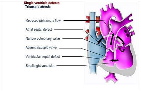
Tricuspid atresia is characterised by the absence of a tricuspid valve and usually a small right ventricle (right lower heart chamber).
With tricuspid atresia (no opening), the valve between the right atrium (upper right heart chamber) and the right ventricle (lower right heart chamber) is missing and there is diminished blood-flow from the right ventricle to the lungs.
Why does this happen and how may it affect the patients health?
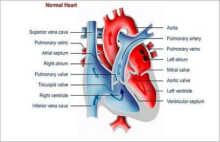
Under normal conditions, the tricuspid valve allows blood to pass from the right atrium to the right ventricle. The blood is then pumped to the lungs where it receives oxygen. It then flows back to the left upper heart chamber (left atrium), followed by flow to the left ventricle (lower left chamber).
With tricuspid atresia, blood cannot reach the lungs and the left side of the heart, unless other defects such as atrial septal defect (ASD) , ventricular septal defect (VSD), or patent ductus arteriosus (PDA) are also present.
Blood-flow to the lungs is possible through the defect that connects the two routes. This results in the mixing of oxygen-poor and oxygen-rich blood at atrial level. The mixed blood is then pumped to the lungs and the rest of the body, which causes the patient to appear cyanotic (blue discoloration of extremities such as the toes, fingers and lips).
Different types of tricuspid atresia exist depending on whether blood-flow to the lungs is obstructed or not.
What symptoms may the patient experience?
The patient appears cyanotic. There is a blue discoloration of extremities of especially the fingers, toes and lips resulting from unoxygenated blood that is pumped into the systemic circulation to the various parts of the body.
How is the diagnosis made and what special investigations are required?
What is the treatment?
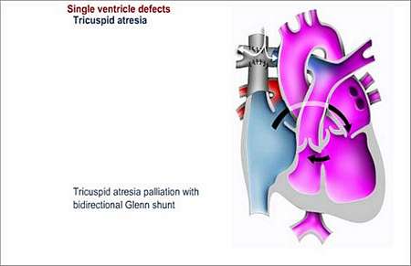
The treatment goal is to improve blood-flow to the lungs by normalising the route of blood-flow as much as possible. A shunt operation is required in the first few months of a babys life. This is a palliative (does not cure the situation, but helps to improve the patients condition) operation in order to increase the amount of blood that flows to the lungs. In patients with unobstructed blood-flow a PA-banding might be required. The types of operations include
- Glenn procedure
A shunt is placed between the superior vena cava (vein that carries deoxygenated blood from the upper body to the heart) and right pulmonary artery. This operation requires the use of a heart-lung machine.
- Fontan procedure
Involves bypassing the right ventricle by joining the right atrium to the pulmonary artery.
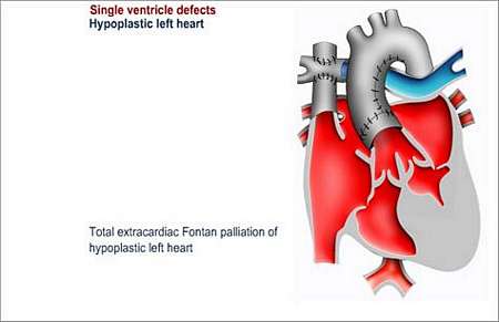
- Total cavo-pulmonary connection
This operation joins the superior and inferior vena cava (vein that carries blood from lower body back to the heart).
Long-term supervision and follow up
It is very important that the patient follow-up with the heart specialist on a regular basis, as no operation corrects the condition completely. Alternatively, patients can visit outpatients for follow-up annually.
Follow-up tests
Preventing endocarditis
The patient will have to take measures to prevent endocarditis for the rest of his or her life. Endocarditis is an infection of the lining of the heart or heart valves.
The patient should wear a Medic Alert card/bracelet stating the need for antibiotics before certain forms of surgery or dentistry.
© 2003 Prometheus™ Healthcare (Pty) Ltd
|